


Scientists at Sanford Burnham Prebys have revealed significant insights into the three-dimensional (3D) structure of the genome and its impact on gene activity. Their research, published on June 27, 2025, in the journal Genome Biology, suggests that the spatial arrangement of DNA within the nucleus plays a crucial role in how genes are regulated, potentially explaining links to various diseases.
Traditionally, the human genome is depicted in a one-dimensional format, which fails to capture the complexity of its structure. Within the nucleus of cells, approximately six feet of DNA is tightly coiled around protein spools known as histones, forming a compact structure called chromatin. This organization is not as random as it appears; the 3D configuration of chromatin features multiple loops and clumps that facilitate interaction among certain genomic regions while isolating others. Disruptions in this 3D structure have been associated with diseases, including developmental disorders and various types of cancer, such as breast cancer and T-cell acute lymphoblastic leukemia.
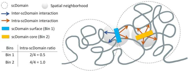
The research team, led by Kelly Yichen Li, Ph.D., a postdoctoral associate at Sanford Burnham Prebys, hypothesized that the 3D shape of genomic regions affects gene regulation. “Many regions of the genome form what are known as topologically associating domains or TADs,” she explained. “Parts of the genome within these domains can interact more frequently with each other, while they tend to be isolated from the regions outside this domain.”
During their spatial mapping experiments, the team observed that TAD-like regions in individual cells often displayed a globular shape. This unique morphology, likened to the irregular contours of potatoes, suggested that these regions could influence the function of nearby genes. Professor Yuk-Lap (Kevin) Yip, Ph.D., the interim director of the Center for Data Sciences at Sanford Burnham Prebys, noted that regions closer to the surface of these chromatin clumps are likely more active due to their exposure to biochemical signals within the cell nucleus.
In their study, the researchers introduced a method to measure how close a genomic region is to the center of a chromatin cluster. “We used a metric to quantify the ‘coreness’ of a genomic region in a chromatin domain,” said Li. Their findings indicate that regions on the surface are more active compared to those buried deep within the chromatin structure.
Yip highlighted the abundant data available for this type of analysis, stating, “The type of data we can apply this measure to is becoming quite plentiful. There is a lot of potential to study how coreness links to gene activity and disease in different cell types.”
The research team plans to collaborate with the lab of Pier Lorenzo Puri, MD, to further investigate the implications of 3D genome structure on muscle stem cell development and the progression of muscular dystrophy. This ongoing research aims to enhance our understanding of how spatial organization within the genome contributes to health and disease.
More information can be found in the study titled “Regulatory roles of three-dimensional structures of chromatin domains” in Genome Biology.