A groundbreaking technique has emerged, allowing scientists to enhance atomic imaging by turning noise into valuable data. This innovative method, known as stochastic Stimulated X-ray Raman Scattering (s-SXRS), offers unprecedented detail in understanding chemical reactions and material properties. The findings of this research were published on July 16, 2025, in the journal Nature.
This advancement was developed by an international team of researchers utilizing the European X-ray Free Electron Laser (XFEL) located in Schenefeld, Germany. Recognized as the largest X-ray laser in the world, the facility enabled researchers to capture detailed snapshots of atomic interactions with remarkable precision. The technique represents a significant leap forward in atomic-scale imaging and could revolutionize the field of chemical analysis.
The research team includes scientists from the U.S. Department of Energy’s Argonne National Laboratory, the Max Planck Institute for Nuclear Physics, and the Small Quantum Systems (SQS) instrument at the European XFEL. Their collaboration has resulted in a method that dramatically improves the resolution of X-ray spectroscopy, which studies electron placement around atomic centers.
“For a long time, chemists have dreamed of observing how electrons move in excited states, as these movements drive chemical reactions,” said Linda Young, an Argonne Distinguished Fellow and professor at the University of Chicago. “Our technique brings us closer to realizing that dream.”
The key innovation of this method lies in its ability to provide super-resolution in X-ray spectroscopy. This advancement allows scientists to identify closely spaced energy levels in atoms, offering clearer insights into their electronic structures, which ultimately dictate chemical properties. Young likened the improvement to upgrading from standard definition to ultra-high definition television, stating, “We’re now able to see the fine details of electronic motion that were previously blurred or invisible.”
The implications of s-SXRS are vast. By providing insights into the formation and breaking of chemical bonds, the technique enhances our understanding of fundamental processes essential for chemical analysis. This knowledge could lead to the development of new materials with specific electronic properties, impacting industries such as electronics and nanotechnology.
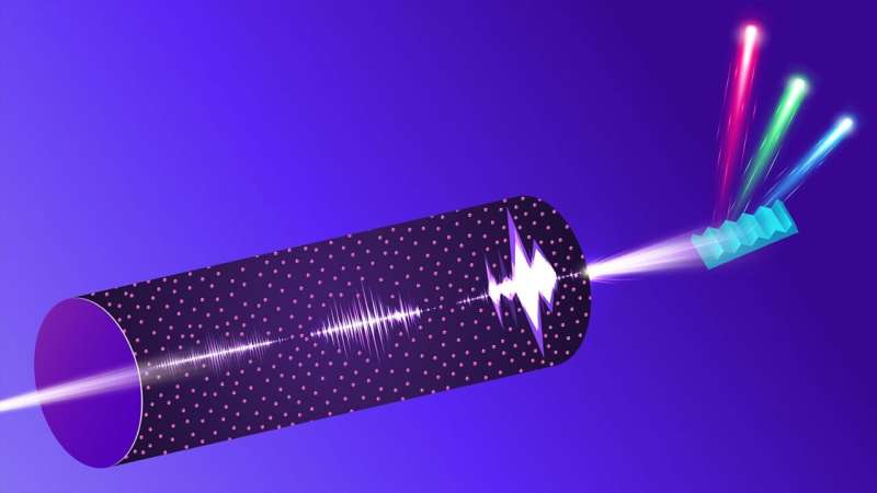
In their experiments, researchers directed X-ray pulses from the European XFEL through neon gas, utilizing a spectrometer designed by the Max Planck Institute for Nuclear Physics. The small, 5-millimeter gas cell facilitated the passage of X-rays, creating tiny openings in the cell’s entrance and exit windows. A grating spectrometer, provided by collaborators from Uppsala University in Sweden, was used to separate light into its various wavelengths.
Experts from the European XFEL played a critical role in coordinating the experimental setup and conducting thorough pre-experimental testing. “This ensured optimal focusing conditions, crucial for efficiently acquiring a large amount of data during the experiment,” explained Michael Meyer, group head of the SQS instrument at European XFEL.
As X-rays traversed the gas, they amplified Raman signals—essentially X-ray fingerprints that reveal information about excited electronic states—by nearly one billion times. This amplified signal grants scientists detailed insights into the electronic structure of the gas on a femtosecond timescale, or one quadrillionth of a second.
By analyzing the relationship between incoming pulses and the resulting Raman signals, researchers can compile a detailed energy spectrum from numerous individual snapshots. This process contrasts with traditional methods that require slow scans across various energy levels.
“The large number of pulses in each X-ray flash boosts the measurement signal and is key to achieving the highest spectral resolution by averaging multiple photon impacts on the detector simultaneously,” noted Thomas Pfeifer from the Max Planck Institute for Nuclear Physics. He further emphasized that the approach mirrors the super-resolution microscopy technique that garnered the 2014 Nobel Prize in Chemistry.
The stochastic Stimulated X-ray Raman Scattering employs a statistical method known as covariance analysis to connect incoming X-ray pulses with emitted Raman signals. This approach transforms previously considered “noise” into a valuable resource, allowing detailed information extraction from complex data. It not only enhances resolution but also accelerates data collection, providing rapid and precise snapshots of atomic interactions.
Another crucial aspect of this research was the involvement of the Argonne Leadership Computing Facility (ALCF), a user facility of the Department of Energy’s Office of Science. The ALCF provided the computational power necessary to simulate the intricate interactions between X-ray pulses and matter. These simulations were vital for interpreting experimental results and refining the technique, closely matching the experimental data.
The continued advancements in s-SXRS could establish it as a standard tool in laboratories worldwide, driving innovation across various scientific fields. Meyer expressed optimism, stating, “This study exemplifies the capabilities of the European XFEL, particularly its high intensities and the generation of extremely short X-ray pulses with durations of less than one femtosecond. These advancements will undoubtedly stimulate further investigations to reveal the dynamics of complex chemical reactions.”
“We’re just beginning to scratch the surface of what we can achieve with this level of detail,” concluded Young. “It’s an exciting time for science and technology.”


