


Researchers at the **Barcelona Institute of Science and Technology (BIST)** have achieved a groundbreaking milestone by filming the exact moment a human embryo implants into the uterine wall. This significant advancement opens new avenues for understanding early human development and may enhance fertility treatments. The ability to capture this crucial step, which previous studies could only depict in snapshots, has been described as a key breakthrough in reproductive science.
Implantation, the process where an embryo attaches itself to the uterine lining, occurs approximately **five to six days** after fertilization. During this time, the embryo is a cluster of **100 to 200 cells**, too small to be detected by ultrasound. Until now, researchers had limited visibility into this critical phase, as it typically happens deep within the uterus. The new experimental setup allows scientists to observe the implantation process dynamically, providing essential insights into an event that can significantly impact pregnancy outcomes.
Revolutionizing Our Understanding of Implantation
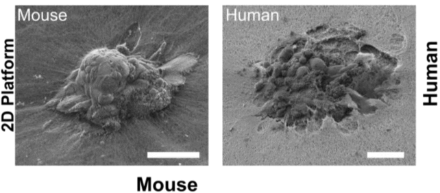
The research team, led by senior author and bioengineer **Samuel Ojosnegros**, developed a specialized platform that recreates the structural environment and nutrient conditions necessary for embryo implantation. This innovative system enabled them to visualize how human embryos interact with a **collagen-based matrix** designed to mimic the uterine environment. In their experiments, they discovered that human embryos aggressively penetrate this matrix, forming a cavity for growth.
“For the first time, we’ve been able to watch human embryo implantation unfold dynamically,” Ojosnegros stated. “We’ve opened a window into a stage of development that was previously hidden.” This technique highlights the invasive nature of human embryos, which exhibit a much deeper penetration compared to mouse embryos, which only traverse the matrix superficially. The researchers noted that understanding this mechanical process is crucial, as approximately **60 percent** of failed pregnancies occur during implantation or shortly thereafter.
The findings suggest that the unique behaviors of human embryos could not be fully understood through studies involving animal models. “Our technology lets us pinpoint where the embryo exerts force,” Ojosnegros added, underscoring the limitations of mouse studies in capturing the complexities of human implantation.
Potential Implications for Fertility Treatments
The implications of this research extend beyond academic curiosity; they may lead to practical applications in fertility treatments. As Ojosnegros explained, the collagen-based matrix is not derived from human uterine cells, which allows for manipulation of its composition to assess how embryos respond to various environments. This flexibility could help identify compounds that enhance implantation success.
Through a collaboration with the pharmaceutical company **Grifols**, Ojosnegros and his team have already begun developing a protein supplement designed to improve implantation rates in clinical settings. Their spin-off company, **Serabiotics**, aims to translate these findings into real-world solutions for individuals facing fertility challenges.
The study, which provides a detailed look at the implantation process, was published in **Science Advances**. By illuminating this critical stage of human development, researchers hope to pave the way for improved fertility interventions and a deeper understanding of early human life.