


In a groundbreaking study, researchers have demonstrated the potential of using photoacoustic microscopy to image stents through the skin. This innovative approach could provide a safer and noninvasive method for monitoring these crucial medical devices, which are implanted in approximately 2 million people each year in the United States to enhance blood flow in narrowed or blocked arteries.
The study, published on July 26, 2025, in the journal Optics Letters, highlights the work of co-lead researchers Myeongsu Seong from Xi’an Jiaotong-Liverpool University and Sung-Liang Chen from Shanghai Jiao Tong University. Traditionally, monitoring stent integrity for issues like fractures or improper positioning has required invasive procedures or exposure to radiation. Seong emphasized the necessity for improved monitoring techniques, stating, “This inspired us to test the potential of using photoacoustic imaging for monitoring stents through the skin.”
Advancements in Photoacoustic Imaging
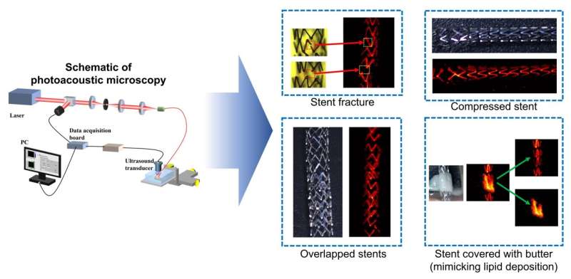
Photoacoustic imaging is an innovative, label-free technique that captures sound waves produced when materials absorb light and release energy. This method offers higher-resolution images at greater depths compared to traditional optical methods, as sound scatters less than light. While previous studies have employed photoacoustic imaging via an endoscope, this still necessitated an invasive procedure for patients.
In their research, the team simulated various conditions affecting stents, such as fractures and plaque buildup, by using butter to replicate lipid deposits or blood clots post-stenting. By utilizing photoacoustic microscopy at different wavelengths, including 670 nm and 1210 nm, they successfully imaged these scenarios through excised mouse skin. Seong noted, “One of the most interesting results is that we could easily differentiate between the butter we used to mimic a lipid plaque and the stent.”
Potential for Clinical Application
The implications of this research could significantly enhance patient care, especially for stents placed in dialysis access sites, which are generally located just beneath the skin. For stents situated deeper, such as those in the carotid artery, a related technique known as photoacoustic computed tomography may be more effective.
Although the results are promising, the researchers acknowledge that further development is necessary before photoacoustic imaging can be utilized in clinical settings. This includes conducting in vivo animal experiments and initial clinical trials to assess its efficacy and optimizing the system for different anatomical locations.
The findings from this study could lead to a revolution in how healthcare professionals monitor stent conditions, offering a noninvasive option that could ease patient anxiety and enhance safety. As Seong concluded, “This would make it easier and safer to monitor the condition of stents in patients.”
For further details, refer to the full study: Siqi Liang et al, “Photoacoustic microscopy for visualization of stents in multiple scenarios,” published in Optics Letters (2025). DOI: 10.1364/OL.564778.